Neuronal Plasticity can be defined as the ability of the nervous system to change structure, function and organization of neurons in response to new experiences, emerging from changes of developmental and environmental situations, as well as other factors (e.g. injuries) affecting the conditions of the nervous system or the whole organism. Neuronal plasticity such as Neurogenesis, Synaptogenesis and Neurite Outgrowth once thought to occur mainly during development has now been recognized as key features of adult brain function.
The balance between neurogenesis and cell death plays a critical role not only in the embryonic brain development but also in the maintenance of the adult brain. Alterations in these processes are seen in many neurodegenerative diseases. The hippocampus, a brain area critical for learning and memory, is especially vulnerable to damage at early stages of Alzheimer’s disease (AD). Emerging evidence has indicated that compromised neurogenesis in the adult hippocampus represents an early critical event in the course of AD. For this purpose, QPS Austria provides a cell culture model to assess effects of your developmental compound on neurogenesis of hippocampal neurons.
Neurogenesis Solution
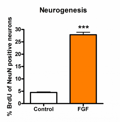
Primary rat hippocampal neurons are treated with compounds on DIV1 for a total of 48 h. After 24 h, BrdU [10 µM] is added. Compounds are present throughout the assay. On DIV3, after 48h incubation cells are subjected to indirect immunofluorescence analysis. Digital images from primary rat hippocampal neurons are analyzed for the percentage of BrdU positive cells compared to the total number of neurons (NeuN positive cells) using a software-supported automatic quantification method. Analysis is performed using Image Pro Plus software. Approximately 10000 to 15000 cells per group are screened for BrdU positive nuclei.

Figure: Effects of Fibroblast Growth Factor (FGF) on neurogenesis of primary rat hippocampal neurons.
Synapse formation but especially the synaptic maintenance (synaptogenesis) is critical for neuronal health and disease. Loss of synaptic maintenance is not only restricted to AD. Defects in synaptic maintenance most likely also contribute to the synaptic vulnerability in other neurodegenerative disorders as Huntington’s, Parkinson’s or Niemann Pick disease. A therapeutic strategy that favors synaptic maintenance may be a beneficial target for many neurodegenerative diseases. For this purpose, QPS Austria provides a cell culture model to assess effects of your developmental compound on synaptogenesis primary hippocampal neurons.
Synaptogenesis Solution
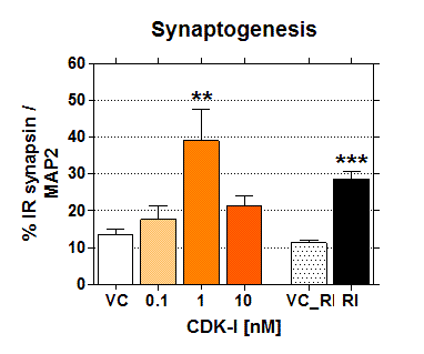
To evaluate effects of Test Item (a commercially available CDK inhibitor) and Reference Item (RI) on synaptogenesis in primary rat hippocampal neurons, cells are treated on DIV6 for 24h. Cells are subjected to immunofluorescence analysis using synapsin and MAP2. Nine systematic uniform random frames are imaged per coverslip at 63x magnification as 10 image deep z-stacks on an AxioImager.Z1 microscope equipped with LED illumination and a Axio MRm B&W camera with 1x opto-coupler. The MAP2 image served to delineate the cells. The area of interest is limited to the neurites of the cells. Results are shown as % immunoreactive area of synapsin co-localization to MAP2 area.

Figure: Effects of Test and Reference Item (RI) on synaptogenesis in primary rat hippocampal neurons.
During development, neurons become assembled into functional networks by growing out axons and dendrites (collectively neurite outgrowth) that connect synaptically to other neurons. Strikingly, neurons retain their capacity for growth and synapse formation in the adult brain. Only those neurons that manage to establish adequate contacts with neighbor cells escape cell death. To stimulate and accelerate neurite outgrowth is thus important for restoring neuronal function after injury or from neurodegenerative diseases. A promising strategy for drugs targeted against neurodegenerative disorders such as Alzheimer’s and Parkinson’s disease is promoting the regeneration of neurons. Thus, new therapies are focusing on identifying new molecules that affect neurite outgrowth. For this purpose, QPS Austria provides cell culture models to assess the neurotrophic effects of your developmental compounds.
Neurite Outgrowth Solution 1
Primary rat (mouse) hippocampal or cortex neurons are treated with compounds for 48 h on DIV1. Cells are subjected to indirect immunofluorescence analyis using anti-beta-III tubulin antibody. The following parameters using image based analysis (Axio.Imager Z1 microscope (20x), 10 image deep z-stacks, collapsed to 2D (ImageProPlus), 12 images per slide from 12 predefined fields, Image enhancement, filtering) are determined of approximately 200 cells per experimental group.
- number of neurites
- total length of neurites
- length of the longest neurite

Figure: Effects of Compound X and reference item (RI) on neurite outgrowth of primary rat hippocampal neurons.
Neurite Outgrowth Solution 2
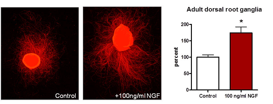
Adult rat dorsal root ganglion (DRG) explants are cultured under serum free conditions in the presence or abesence of neurotrophic factors for 5 days. Explants are are subjected to indirect immunofluorescence analyis using anti-beta-III tubulin antibody. Parameters such as the neuritic halo (the total area specifically occupied by neurites) are analyzed by using a software-supported automatic quantification method.

Figure 2: Neuritic halo of adult dorsal root ganglion explants upon treatment with NGF.
