Multi-Channel Imaging to Visualize Renal Inflammation in an Alport Disease Mouse Model
While we are specialists for neuronal tissue, we also routinely process tissue of peripheral organs:
Kidney of the Alport syndrome mouse model Col4A3-/- was labeled for macrophages and leukocytes (Fig.1) as well as T-lymphocytes and macrophages (Fig.2), followed by automated slide scanning.
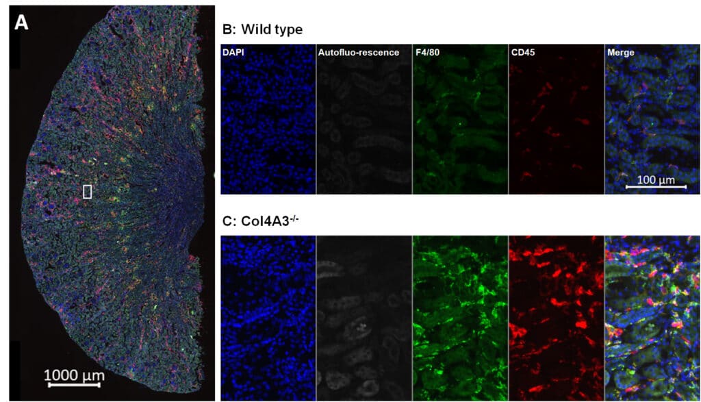
Figure 1: Immunofluorescent labeling for macrophages (F4/80) and leukocytes (CD45). A: whole kidney. B and C: detail of A in wild type (B) and Col4A3-/- mice (C).
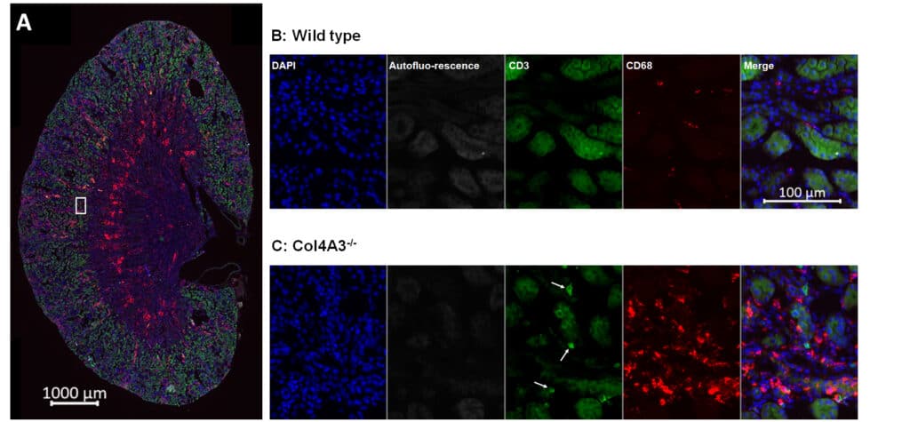
Figure 2: Immunofluorescent labeling for T-lymphocytes (CD3) and macrophages (CD68). A: whole kidney. B and C: detail of A in wild type (B) and Col4A3-/- mice (C).
You can’t find the time to perform your histological experiments?
Think about outsourcing to our experts!
Embedding
- Provide your fixed samples and we embed them for you
- RNA-free procedures are available
Sectioning & Collecting
- Sectioning on cryotome or vibratome
- Choose orientation, thickness, sampling scheme
Immunohistology & General Stainings
- 4-channel immunofluorescence plus DAPI labeling of nuclei
- Hundreds of established antibodies
Microscopy & Imaging
- Up to 5-channel epifluorescence slide scanning
- Appropriate imaging controls
Quantitative Image Analysis
- Identification and detection of labeled structures
- Analysis of region and object features
Advantages of outsourcing your histological experiments to QPS Neuropharmacology
- Improve the quality of your labelings and stainings
- Improve your study efficacy
- Increase your flexibility
- Save time and money
- Reduce your risks